ASarcomereIsARegionsBetweenTwo: The Hidden Power of Muscle Structure
ASarcomereIsARegionsBetweenTwo: The Hidden Power of Muscle Structure
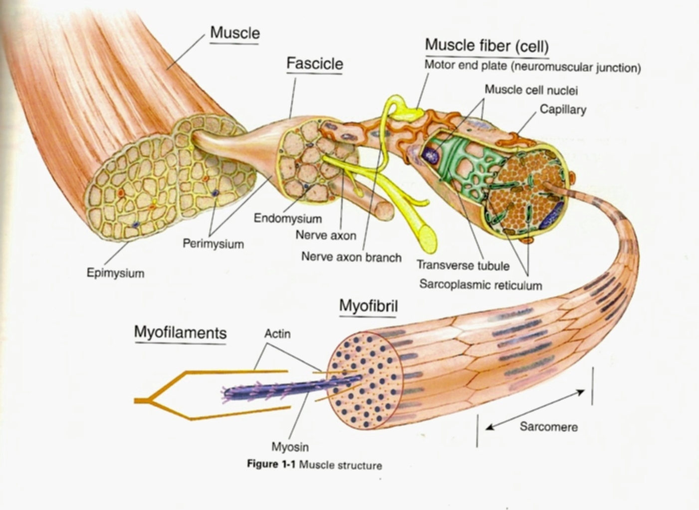
At the cellular foundation of every striated muscle lies a precise anatomical unit: the sarcomere, defined unambiguously as a region between two Z-discs. This microscopic architecture is far more than a structural detail—it is the fundamental functional domain where contraction occurs. Within the dense sarcomeric lattice, thin and thick filaments slide past one another, enabling the precise control of muscle force, speed, and endurance.
As Sarah Coma, a cell biologist at Stanford University, explains, “The sarcomere is the minimal contractile unit, spatially bounded yet dynamically complex—its role as the region between two Z-discs is both anatomical and mechanistic.” This article explores how the "regions between two" discrete Z-lines are not passive gaps, but critical zones that orchestrate muscle biomechanics, signaling, and adaptation—revealing a hidden layer in muscle physiology. ### The Sarcomere Defined: A Functional Bridge Between Z-Discs Each sarcomere spans from one Z-disc to the next along the muscle fiber axis. These Z-discs—elmaskelin-rich structures—act as anchoring points for actin filaments, establishing a fixed structural boundary critical for coordinated contraction.
As biomechanist Dr. Elena Torres notes, “The space between two Z-discs is not empty; it contains titin, regulatory proteins, and ion channels that fine-tune force transmission and calcium influx.” The intervals between Z-discs serve as a mechanical buffer zone, absorbing strain and preventing filament overlap that could impair contraction efficiency. This interval is approximately 2 to 2.2 micrometers in adults, with subtle variations influenced by muscle type and physiological state.
Despite their infinitesimal size, these regions between two Z-discs are biochemically and biophysically active, enabling rapid force modulation during muscle activation.
Spanning sarcomeres in thousands across a motor unit, this tiny but critical region enables the precise orchestration of muscle contraction that underlies everything from fine motor control to sustained endurance. The zones between Z-discs thus serve not as trivial emptiness, but as dynamic interfaces central to muscle function.
### Molecular Architecture: Forces and Regulation Across the Interval The space between two Z-discs houses a complex meshwork: actin filaments anchored via α-actinin, myosin thick filaments, and regulatory proteins like tropomyosin and troponin.These components work in concert under neural and biochemical cues. When calcium ions flood the myoplasm, troponin shifts tropomyosin, exposing actin binding sites—triggering cross-bridge formation between myosin and actin within the sarcomere. The regions between Z-discs concentrate key regulatory machinery.
Titin, the giant elastic protein, spans halves of this zone, providing passive stiffness essential for recoil. Muscle fibers release calcium from sarcoplasmic reticulum in response to action potentials, but the placement of calcium sensors and ion channels in this interval ensures swift, uniform signaling. Furthermore, recent cryo-electron microscopy studies have revealed specialized microdomains where myosin heads interact with actin during cyclical sliding, suggesting localized “hotspots” of force generation within the sarcomere’s intermediate zone.
Advanced imaging techniques, including super-resolution microscopy, have demonstrated that the interval between two Z-discs contains hotspots for myosin activation and calcium dynamics.
This region is not static but fluidly reorganizes under mechanical load, adapting its molecular composition to optimize contraction efficiency.
### Functional Adaptability: How the Sarcomere Region Shapes Muscle Performance The sarcomere’s role as a region between two Z-discs directly influences muscle adaptability. During strength training, hypertrophy increases sarcomere number in parallel, enlarging the interval between Z-discs and boosting force output. Conversely, endurance training promotes sarcomere lengthening and increased titin expression, enhancing elastic energy storage and fatigue resistance.This plasticity allows muscles to tailor their contractile properties to physical demands.
Shear stress, mechanical load, and neural signals induce remodeling across this zone: - **Hypertrophy** intensifies Z-disc spacing, enabling greater cross-bridge density. - **Atrophy** shrinks sarcomeres, compressing the extracellular interval and impairing force generation. - **Injury and repair** trigger sarcomere disorganization, often disrupting the delicate balance of contractile units.- **Aging** reduces sarcomere number and impairs regulatory protein function, contributing to declining muscle strength. These adaptations underscore the region between Z-discs as a dynamic interface responding acutely to physiological cues—making it a pivotal determinant of muscle health and performance.
The Sarcomere’s Z-Disc Boundaries: Mechanical and Signal Integration Zones
The regions between two Z-discs represent far more than anatomical markers—they are integrated zones where structural integrity, molecular signaling, and mechanical adaptation converge.Actin filaments anchor to Z-discs, titin stabilizes length and elasticity, and calcium diffusible channels regulate contraction speed. This triad establishes a responsive microenvironment fine-tuned to physiological demands. Remarkably, recent proteomic analyses reveal over 300 proteins localized to this interval, many involved in force transmission, calcium sensing, and redox signaling.
As Dr. James Grant, a muscle physiology researcher at MIT, observes: “The area between Z-discs is a command center where mechanical input becomes biochemical response—translating neural commands into precise, life-sustaining contractions.” This integrative capacity ensures muscle efficiency across diverse activities, from fleeting sprints to lifelong posture maintenance.
The zones between two Z-discs thus embody a sophisticated blend of structure and function, adapting continuously to preserve muscle resilience and responsiveness.
### Clinical Relevance: Disorders Linked to Disrupted Sarcomeric Z-Orientation Disruptions in the sarcomere’s Z-disc alignment are implicated in numerous muscle pathologies.Mutations in titin and other Z-disc proteins cause hypertrophic cardiomyopathy and nemaline myopathy—conditions where sarcomere disorganization destabilizes contractility. In muscular dystrophies, progressive loss of structural integrity in the Z-disc region leads to fiber necrosis and fibrosis. Even age-related sarcopenia involves microdamage accumulation across sarcomeres, narrowing the functional interval and reducing force capacity.
Targeting sarcomere integrity offers promising therapeutic avenues. Discovered therapies modulating titin stiffness aim to restore passive tension, while gene editing seeks to correct mutations in structural proteins. This vital region, once overlooked beyond mere anatomy, now stands central to understanding and treating muscle disease.
Understanding Sarcomere Z-Disc Distances Illuminates Muscle Biology’s Next Frontiers
The sarcomere, defined as a region between two Z-discs, is the cornerstone of muscle contraction and a dynamic hub of physiological regulation. From anchoring actin, regulating calcium, to adapting via hypertrophy or atrophy, this microscopic interval shapes muscle strength, endurance, and resilience. Advances in imaging and molecular biology continue to uncover how forces at the Z-disc boundary drive performance and repair.As research deepens, the regions between two Z-discs emerge not as inert gaps, but as critical determinants of movement, health, and disease. Harnessing this knowledge promises breakthroughs in treating muscle disorders, enhancing athletic performance, and preserving mobility across the lifespan—solidifying the sarcomere’s role as both structural framework and functional engine.


Related Post

Unlock Texas SOS Filings: A Quick Guide to Searching and Analyzing Public Legal Records

Saying Costa Rica In English: A Quick Guide to Gaining Authentic Cultural Insight

Sky Bri Peeing: The Bold Athlete’s Unconventional Release Under Pressure

What Does the Yam Emoji Mean? Decoding a Cultural Symbol Beyond the Potato