Behind the Rhythm: The Unique Features of the Human Heart
Behind the Rhythm: The Unique Features of the Human Heart
The heart, a muscular marvel beating tirelessly since birth, orchestrates circulation through exquisite anatomical design and physiological precision. As the central pump of the cardiovascular system, it features a complex, compartmental structure optimized for blood propulsion with unmatched efficiency. From its two chambers that inaugurate blood flow to its powerful muscular walls that generate relentless contractions, the heart’s design reflects millions of years of evolutionary refinement.
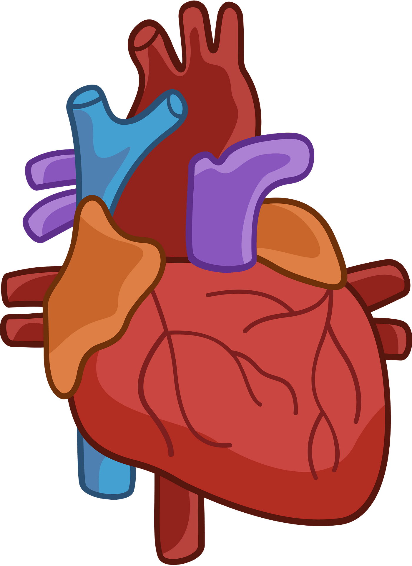
Understanding its key features—chambers, valves, conduction system, and vascular connections—reveals not only its mechanical genius but also its critical role in sustaining life. The heart is conventionally divided into four distinct chambers: two atria and two ventricles, each fulfilling a specialized function in the circulatory circuit. The right atrium receives deoxygenated blood from the systemic circulation via the superior and inferior vena cavae, while the left atrium collects oxygen-rich blood returning from the lungs through the pulmonary veins.
This segregation ensures efficient oxygenation and prevents arterial circulation of deoxygenated blood. The ventricles, more muscular and structurally robust, actualize this separation through powerful contractions. The right ventricle pumps deoxygenated blood into the pulmonary artery en route to the lungs, whereas the left ventricle—possessed of the thickest myocardial wall—forces oxygenated blood into the aorta, driving systemic perfusion.
Integral to this function are the heart’s four essential valves, each engineered to maintain unidirectional blood flow and prevent backflow. The tricuspid valve separates right atrium and ventricle, opening during diastole to allow inflow and closing sharply during systole to block regurgitation. Similarly, the bicuspid (または mitral) valve guards the left side of the heart, separating the left atrium from the left ventricle with identical precision.
Atrioventricular (AV) valves in this configuration respond dynamically to pressure changes, contracting gently to support forward flow. During the cardiac cycle, these valves open in sync with pressure differentials, closing with a distinctive "lub" sound, a clinical hallmark recognized since auscultation began centuries ago. "The valves are the guardians of direction," explains cardiologist Dr.
Elena Marquez, emphasizing their role in maintaining hemodynamic stability.
The heart’s propulsion system hinges on a coordinated electrical conduction network, beginning at the sinoatrial (SA) node—the heart’s natural pacemaker—located in the right atrium. Electrical impulses originating here spread rapidly across atrial muscle, triggering contraction and pushing blood into the ventricles. From the atria, signals travel through the atrioventricular (AV) node, briefly pausing at the internodal region to allow ventricular filling before advancing to the bundle of His and Purkinje fibers.
This pathway ensures synchronized myocardial contraction, with ventricular activation beginning at the apex and spreading upward in a precisely timed sequence. This electrical-mechanical coupling is fundamental to the heart’s rhythmicity and efficiency, accounting for the steady 60–100 beats per minute observed in healthy adults. Disruptions to this conduction system underlie arrhythmias, illustrating how delicate the electrical fine-tuning truly is.
The heart’s inner lining—endocardium—is a smooth, endothelial layer that reduces friction as blood flows through chambers and vessels. Beneath lies the muscular myocardium, composed of cardiac muscle cells uniquely adapted for continuous contraction without fatigue. These cells contain intercalated discs—specialized junctions that link adjacent myocytes via gap junctions and desmosomes—enabling rapid electrical transmission and mechanical cohesion.
The myocardium’s thickness varies markedly: the right ventricle is relatively thin, suited for low-pressure pulmonary circulation, while the left ventricle’s thick, dense walls generate the high pressure required to supply the entire body. This regional variation exemplifies functional optimization, ensuring each segment performs under its specific physiological demands.
Supplying and draining the heart require an exquisitely tailored vascular network.
Coronary arteries branch directly from the ascending aorta just above the aortic valve, delivering oxygen and nutrients essential for sustained contraction. Notably, these vessels supply only the myocardium—no middle layer of coronary vasculature supports the heart wall indirectly—making coronary perfusion pressure a critical factor in cardiac health. Intertwined with this arterial supply is the venous return system: deoxygenated blood from the heart drains via the pulmonary veins into the left atrium, and oxygen-depleted systemic blood returns through the superior and inferior venae cavae.
This closed loop ensures uninterrupted circulation, with the heart’s valves preventing backflow at every junction.
Beyond structure and function, the heart’s resilience and adaptability underscore its complexity. It adjusts output dynamically—doubling cardiac output during exercise by increasing stroke volume and heart rate, driven by autonomic regulation.
The heart also exhibits limited regenerative capacity, highlighting clinical challenges in repair following injury. Yet, advances in imaging, electrophysiology, and surgical repair continue to deepen understanding and restore function in damaged tissue. The heart’s four-chambered architecture, coupled with its sophisticated conduction, valve control, and vascular integration, forms a system so precisely engineered that its failure often spells crisis.
In essence, the heart’s features—chambers defining flow paths, valves ensuring orderly propulsion, a synchronized conduction system maintaining rhythm, and specialized tissues supporting endurance—collectively define its role as the body’s lifeline. Each component, though distinct in form, contributes to an integrated whole that operates with astonishing precision. Mastery of these features is not only vital for medical science but essential for empowering individuals to appreciate and protect this irreplaceable organ.


Related Post

Define Robber Baron: How Visionary Dominance Shapes Modern Power

Master Instagram Nip Slip: The Revolutionary Technique Reshaping Digital Fashion & Self-Expression

Liverpool vs. United 2009: When Titans Collide in a Defining English Derby

Everything You Need to Know About OnlyFans Pages: A Comprehensive Guide