Inside the Petrous Bone: The Hidden Key to the Brain’s Most Fragile Nerve
Inside the Petrous Bone: The Hidden Key to the Brain’s Most Fragile Nerve
The petrous bone, a dense, pyramid-shaped segment of the temporal bone, stands as one of the most critical yet underrecognized structures in human anatomy—serving as both a fortress for the brainstem and the labyrinthine passageway for the facial nerve, among other vital neural and vascular conduits. Nestled at the base of the skull, this compact but powerful bone forms the posterior-most portion of the temporal bone, enveloping the internal auditory meatus—a narrow canal guarding the delicate nerve basilics of cranial nerve VII. Far more than a mere protective casing, the petrous bone’s intricate architecture enables life-sustaining functions, making it a focal point in neurosurgery, otolaryngology, and neurology.
Its role in shielding the brainstem—the core of vital autonomic control—underscores its surgical and clinical significance, while its central position in the skull’s neurovascular landscape explains why damage or pathology in this region can have profound and lasting consequences.
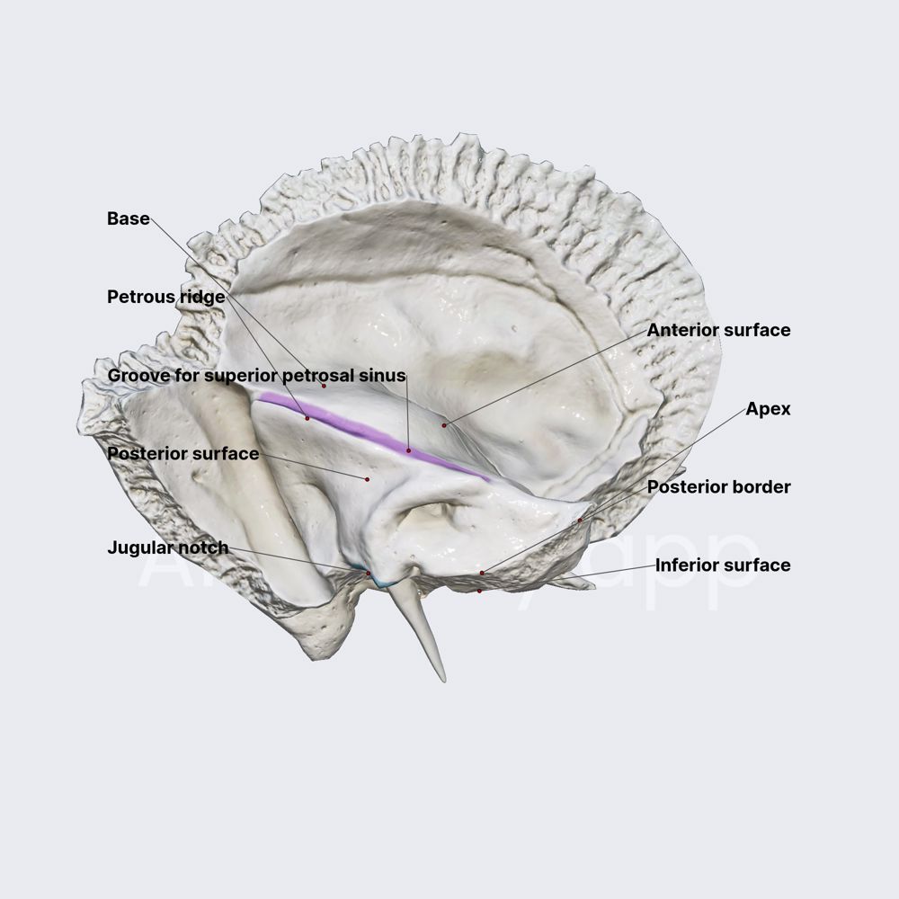
Designed by evolution to fulfill dual protective and transmission roles, the petrous bone is a marvel of biological engineering. Composed primarily of dense cortical bone, it features a convoluted internal architecture designed to house and safeguard several critical structures.
At its core lies the internal auditory canal (or meatus), a rigid channel formed almost entirely within the petrous portion, through which cranial nerves VII (facial), VIII (vestibulocochlear), and part of IX (glossopharyngeal) descend to reach the brainstem. This bony tunnel measures roughly 2.5 centimeters in length but contains remarkable features: microscopic foramina permit passage of nerve fibers and tiny vessels, while trabecular internal architecture evenly distributes mechanical stress, reducing fracture risk at this uniquely vulnerable skull region. “The petrous bone’s design is nothing short of ingenious—protecting the most sensitive neural infrastructure while maintaining structural resilience,” notes Dr.
Elena Morales, a neuroradiologist with decades of experience in skull base imaging.
The petrous bone’s clinical relevance becomes most apparent when considering its central role in protecting the facial nerve (cranial nerve VII), one of the most complex cranial nerves responsible for motor control of facial musculature, taste from the anterior two-thirds of the tongue, and parasympathetic innervation to glands. With over 99% of facial nerve fibers traversing the internal auditory canal, any structural compromise—whether from trauma, tumor, or inflammatory disease—can trigger life-altering deficits, including facial paralysis.
“The petrous bone is practically the facial nerve’s last line of defense,” explains Norman DN, an expert in skull base surgery. “Its integrity is paramount; even subtle erosion or swelling can jeopardize recovery after intervention.” Beyond the facial nerve, cranial nerve VIII—essential for hearing and balance—also originates within this bony vault, making the petrous bone indispensable for auditory and vestibular function. Damage here, whether from tumor growth, infection, or surgical error, typically results in profound hearing loss or vertigo.
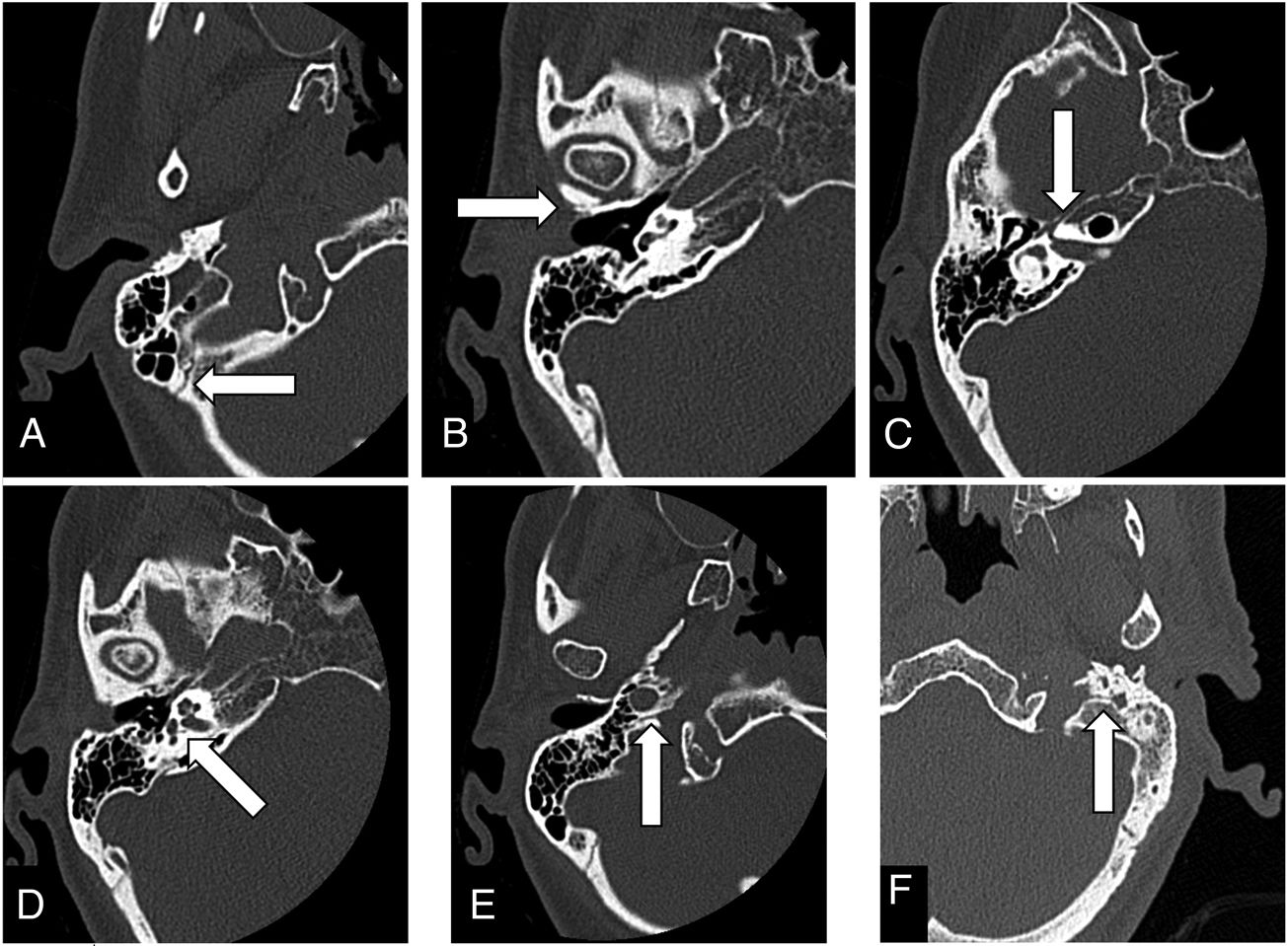
Neurosurgeons and radiologists frequently rely on imaging modalities such as high-resolution computed tomography (CT) and magnetic resonance imaging (MRI) to assess the petrous bone’s condition. These tools reveal not only its bony structure but also surrounding soft tissue dynamics, such as nerve displacement, vascular enlargement, or neoplastic invasion—critical factors in diagnosing conditions like petrous periosteal osteosarcoma, arbitrary skull base meningiomas, or chronic otogenic infections. “In tumor surgery, preserving the petrous bone while ensuring complete tumor resection demands exquisite precision,” warns Dr.
Sarah Patel, a neurosurgeon specializing in temporal fossa procedures. “Modern neurosurgical techniques, including endoscopic endonasal approaches, allow for minimally invasive access with minimal disruption to surrounding neural tissue—showcasing how evolutionary anatomy is merging with cutting-edge intervention.”
The petrous bone’s vulnerability is underscored by its location: perched at the skull base, it lies in close proximity to major vascular structures including the carotid artery, internal carotid artery, and jugular vein—making trauma or pathological expansion here a race against catastrophic neurological compromise. Epidural hematomas, epidural skull fractures, and cerebrospinal fluid leaks often involve the petrous region, emphasizing the need for rapid diagnosis and intervention

Related Post

Christian Brauns Wife Life Love And Family: A Deep Look at Faith, Partnership, and Private Blessings

Decoding Card Formats: The Hidden Language Behind Smart Cards

Kisah Istri Selingkuh: Fakta dan Dampak sejarah claimed sebelum Persatuan Indonesia Nasional crystallize

Brandy Talore: Breaking Barriers in the Adult Film Industry Through Authenticity


