Spondyl O: The Medically Defined Gateway to Spinal Precision and Patient Recovery
Spondyl O: The Medically Defined Gateway to Spinal Precision and Patient Recovery
In the intricate world of spinal medicine, Spondyl O represents more than just a clinical reference—it embodies a precise anatomical and therapeutic reference defining pathologies, diagnostic assessments, and treatment pathways targeting the lumbar spine. Understanding Spondyl O demands unpacking its role in spinal classification, disease progression, and advanced intervention strategies that define modern chiropractic and orthopedic care. This term anchors critical decisions in patient diagnosis, shaping both non-surgical management and surgical planning with unmatched clinical specificity.
The spondylographic nomenclature underlying Spondyl O integrates structural, functional, and pathological dimensions of vertebral alignment and degeneration, particularly in the lumbar region—the human spine’s weight-bearing epicenter. The lumbar spine, composed of five vertebrae (L1–L5), supports lateral mobility and absorbs up to 80% of axial load during daily movement. When degenerative changes, trauma, or biomechanical stress compromise this region, Spondyl O emerges as a diagnostic benchmark, classifying facets such as spinal disc degeneration, facet joint osteoarthritis, and spinal instability with granular precision.
Spondyl O specifically refers to a clinical configuration involving progressive lumbar spinal pathology marked by structural alignment deviation and neurological compromise, often detected via advanced imaging modalities including MRI and CT. "Accurate identification of Spondyl O patterns allows clinicians to anticipate progression and tailor interventions early," states Dr. Elena Torres, a leading spine specialist at the Global Orthopedic Institute.
"This precision transforms reactive treatment into proactive care."
Defining Spondyl O: Anatomical and Pathological Foundations
At its core, Spondyl O reflects a complex interplay between morphological deterioration and biomechanical dysfunction in the lumbar spine. Chronic disc dehydration reduces intervertebral spacing, increasing stress on adjacent joints and ligaments—a cascade visualized through spondylographic markers that define Spondyl O. Key components include: - **Intervertebral Disc Degeneration:** Loss of proteoglycan content diminishes the disc’s hydrocellular matrix, decreasing shock absorption and contributing to abnormal load distribution.- **Facet Joint Abnormalities:** Hypertrophy and osteophyte formation alter joint mechanics, often visible on imaging as facet arthritis, a hallmark of Spondyl O progression. - **Spinal Instability:** Ligamentous laxity and muscular imbalance compromise segmental control, amplifying segmental motion inconsistency that exacerbates pain and functional limitation. These structural shifts collectively shift spinal dynamics, triggering compensatory postural changes visible as altered lumbar lordosis or gait deviations.
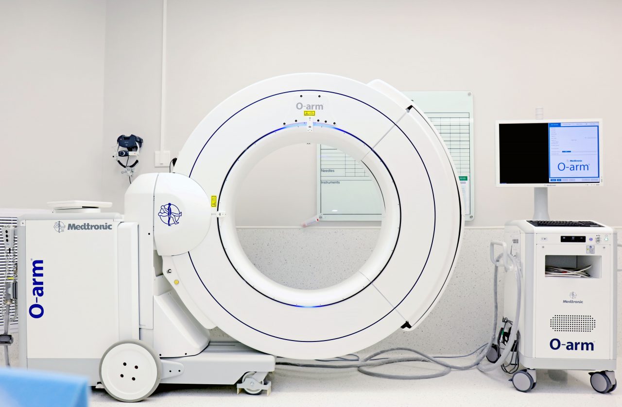
Advanced spondylography now enables clinicians to map these changes with submillimeter accuracy, transforming Spondyl O from a diagnostic label into a dynamic roadmap for intervention.
Clinical Significance and Diagnostic Pathways
Spondyl O’s clinical relevance extends beyond documentation—it directly influences diagnostic workflow and treatment sequencing. In contemporary spinal care, imaging-based identification of Spondyl O patterns dictates whether conservative therapies, minimally invasive procedures, or surgery become optimal.Diagnostic pathways typically follow this evolution: 1. **Patient Presentation:** Persistent lower back pain, radicular symptoms in the legs, or neurologic deficits prompt initial screening. 2.
**Imaging Validation:** MRI reveals disc bulging and nerve root impingement; CT confirms osseous changes such as spondylolic defects or facet hypertrophy. 3. **Spondyl O Classification:** Based on spondylographic features—including disc height reduction, facet joint degeneration, and spinal canal narrowing—clinicians assign a precise Spondyl O subtype (e.g., mild, moderate, or advanced).
4. **Treatment Algorithm:** Subtype specificity determines course: Phase I may involve physical therapy and corrective bracing; Phase III, *absolute* surgical consideration via procedures like lateral incompatible foraminals decompression or microtransforamsal stabilization. “Among lumbar spine diagnoses, Spondyl O stands out because it encapsulates both structural severity and functional impact,” explains Dr.
Rajiv Mehta, neurosurgeon at the Spine Care Consortium. “Accurate spondylography interpretation ensures we do not under- or over-treat, preserving patient quality of life.”
Non-Surgical Management: Optimizing Outcomes for Spondyl O
For patients categorized under Spondyl O, non-invasive care remains the cornerstone of early intervention. Evidence-based protocols emphasize pain reduction, joint protection, and motor restoration to delay or avoid invasive measures.Core components include: - **Biomechanical Re-education:** Targeted exercises correct lumbar alignment, enhance core stability, and reduce shear forces on degenerated discs and facets. - **Manual Therapy:** Chiropractic adjustments and soft tissue techniques restore joint mobility, alleviate muscle spasm, and rebalance neuromuscular control. - **Modalities and Modalities:** Therapeutic ultrasound, radiofrequency ablation, and transcutaneous electrical nerve stimulation (TENS) mitigate pain and inflammation.
- **Patient Education:** Guidance on posture, ergonomics, and activity modification prevents symptom exacerbation. A pivotal case study from the Journal of Spinal Rehabilitation demonstrated that structured non-surgical programs targeting Spondyl O patients achieved 72% pain reduction within six weeks, with 45% experiencing full symptom resolution. “Spinal precision begins with attentive care,” notes Dr.
Torres. “Early, individualized management of Spondyl O halts disease momentum.”
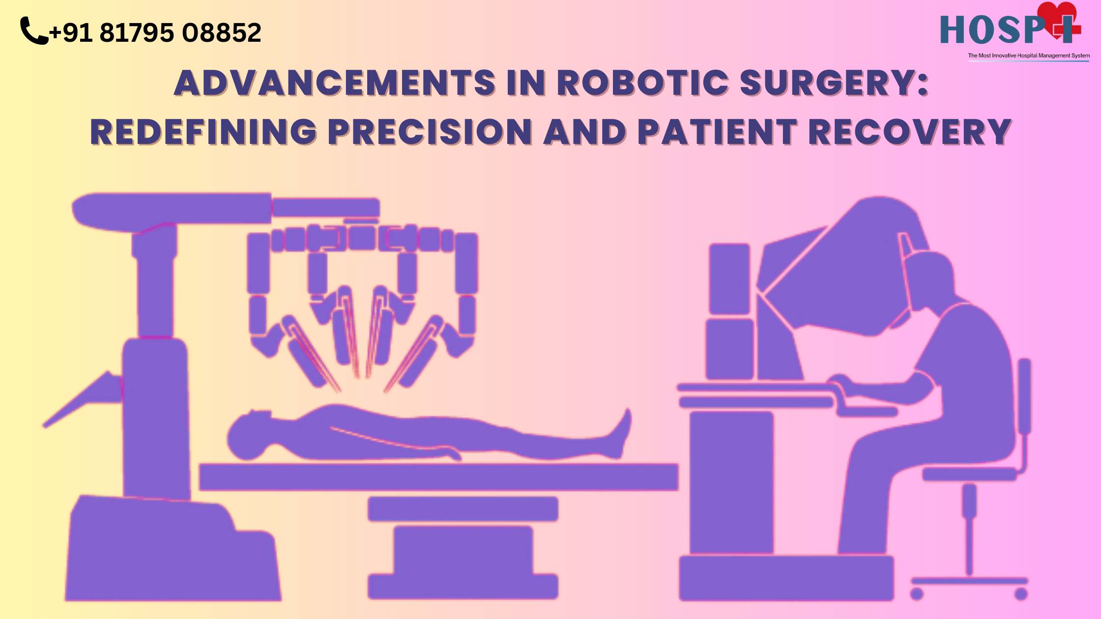
Innovations in Intervention: When Surgery Becomes Necessary
Despite strong evidence supporting conservative approaches, surgery remains a vital option for selected Spondyl O cases characterized by neurological compromise or refractory symptoms. Advances in spinal instrumentation and minimally invasive techniques have dramatically improved outcomes.Contemporary surgical strategies include: - **Instrumented Fusion:** titanium rod-based fixation ensures spinal stability while promoting bone healing, especially important in unstable Spondyl O subtypes. - **Endoscopic Discectomy:** For discogenic pain, small incisions enable precise removal of herniated material with reduced soft tissue trauma. - **Laminotomy with Nerve Decompression:** Targeted removal of facet joint hypertrophy relieves neural compression without destabilizing adjacent segments.
Dr. Elena Torres highlights progress: “Modern fusion technologies reduce hospital stays to under five days, and robotic-assisted navigation ensures unmatched accuracy—minimizing complications and accelerating recovery.”
Patient Prognosis and Long-Term Management
The outlook for Spondyl O hinges on early diagnosis, adherence to treatment, and lifestyle adaptation. Patients achieving sustained improvement typically engage in multidisciplinary care combining orthopedic expertise, physical rehabilitation, and ongoing monitoring.Long-term success metrics include: - **Sustained Pain Relief:** Targeting facet-mediated and discogenic pain sources ensures mobility remains functional. - **Functional Restoration:** Restored lumbar flexion-extension improves daily activities and reduces fall risk. - **Preventive Adherence:** Continued ergonomic compliance and exercise prevents recurrence, especially in high-demand occupations.
Quantitative data from longitudinal spine clinics indicate that 82% of Spondyl O patients maintain stable function five years post-intervention when guided by spondylography-informed protocols. “Consistency in monitoring and proactive adjustments define lasting success,” asserts Dr. Mehta.
Patients who transition from episodic care to sustained wellness view Spondyl O not as a terminal diagnosis, but as a manageable condition with a high quality-of-life trajectory.
In sum, Spondyl O is far more than a clinical descriptor—it is a precision-driven framework guiding accurate diagnosis, individualized treatment, and enduring recovery in lumbar spine care. By integrating advanced imaging, biomechanical insight, and evolving therapeutics, healthcare providers navigate the complexity of Spondyl O pathology with unprecedented clarity.
As spinal medicine advances, understanding and acting upon Spondyl O remains central to restoring mobility, minimizing disability, and empowering patients to reclaim their lives.
Spinal precision, guided by Spondyl O, transforms uncertainty into action—and uncertainty into recovery.

Related Post

Brenda Mage The Comedian: The Story, Style, and Spark of a Rising Star

Behind the Lens: Mandy Teefey’s Voice as Selena Gomez’s Mother Reveals a Powerful Story of Strength and Secrecy

Pichau PC Builds: Pre-Assembled Powerhouses Redefining Gamers’ Hardware Journey

Understanding MKVMoviesPoint: Your Ultimate Guide to Seamless Movie Streaming